© Asia Eye Care Limited. All rights reserved.
In order to determine what is best for the patients, thorough understanding of their ocular condition is required. This is archived through detailed inquiries into their family history of ocular pathology, medication, general health and visual habits.

The visual acuity (sight clarity) and refractive error (myopia, hyperopia, astigmatism or presbyopia) are tested with the retinoscope, ophthalmic lenses and internationally recognized visual acuity charts. Various visual acuity charts (letters, numbers, shapes, pictures or E charts) are utilized for patients of different ages. Children with amblyopia (lazy eyes) are identified using these tests.

Assessment of binocular vision functions (co-ordination of the two eyes) would identify any strabismus (squint) and other extra-ocular muscles problems. Identifying the natures of the strabismus is crucial for effective management.

Ishihara or other age appropriate color vision tests are used to identify color vision deficiency.

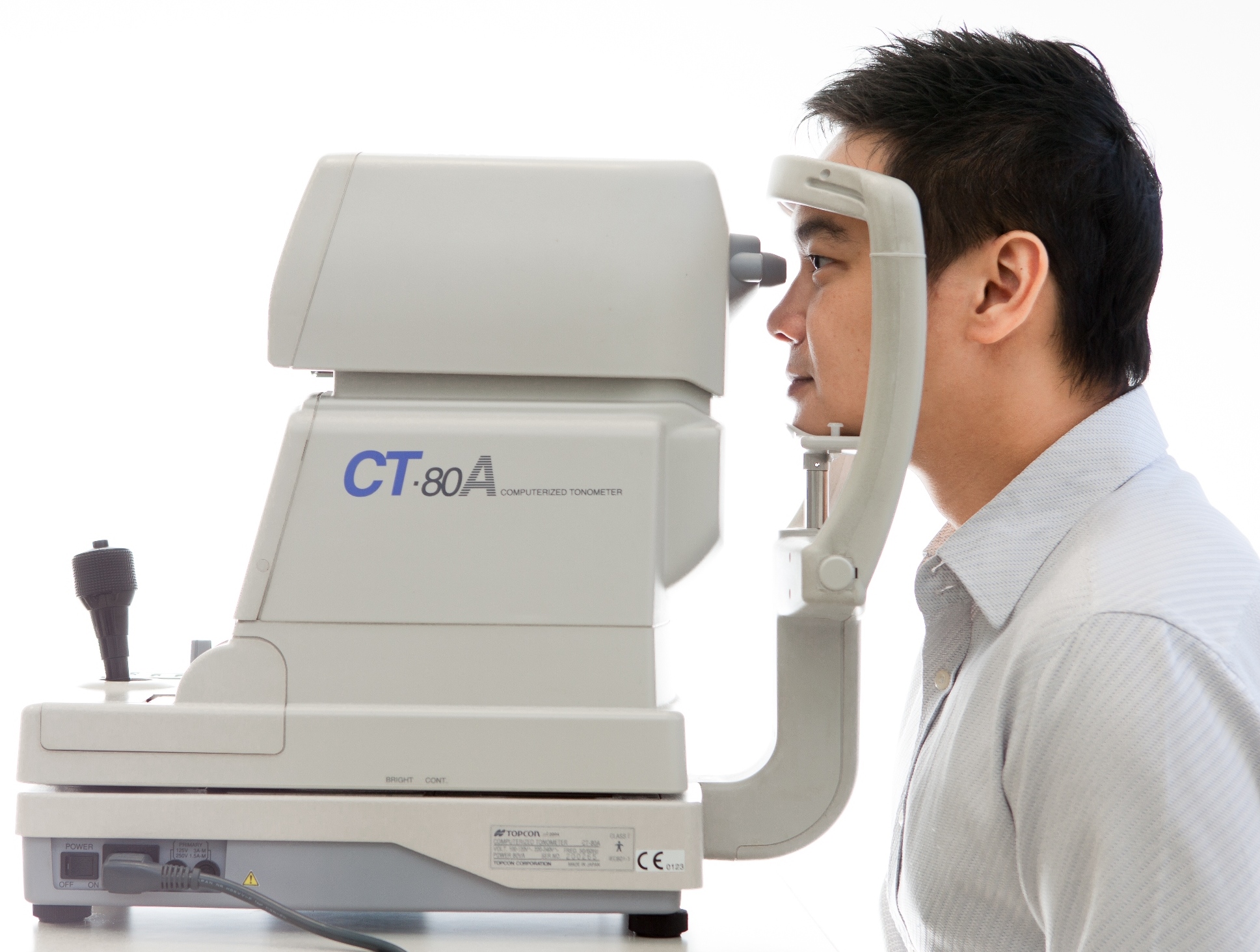
Measurement of the intra-ocular pressure as a preliminary glaucoma assessment.

Comprehensive ocular health assessment on cornea, lens, retina, macula etc. would be carried out by the use of a series of professional ophthalmic equipment, including: slit lamp biomicroscope, ophthalmoscope, binocular indirect ophthalmoscope, fundus camera, funduscopic lenses, tonometer and goniolens. Dilation of the pupils (with mydriatic eye drops) for peripheral retinal examination would be conducted as a routine for most patients. Mydriatic or cycloplegic drops for children would be used on indication.

With the use of high technology fundus camera, the retinal images (including optic nerve, macula, retinal nerve fibers and blood vessels) can be used for diagnostic purposes and for monitoring of any pathological changes.

Our optometrists will review and analyze the patients' ocular condition based on the examination results. Appropriate management and recommendation will be discussed with the patients for their best interests.